Maestro of Microscopy
Published on August 2, 2022New microscopy technology allows scientists to pull more data from samples than ever before
New microscopy technology allows scientists to pull more data from samples than ever before
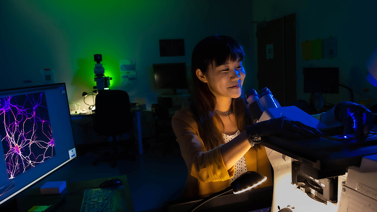
Most people are probably familiar with the small tabletop microscopes they used in their high school biology class. The ones that Michelle Itano ’12 (PhD) works with are called compound light microscopes, which use an eyepiece, set of mirrors and multiple lenses to magnify objects.
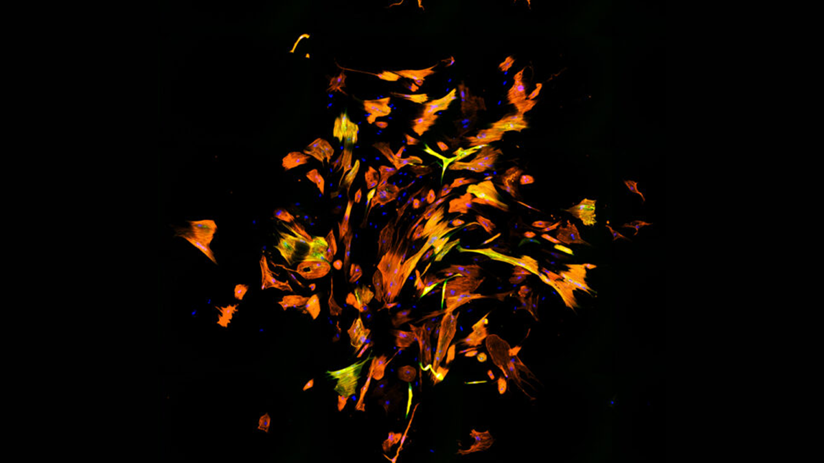
Itano specializes in the fluorescence microscope, a light microscope that uses various wavelengths — or colors — of light that interact with dyes. Users “paint” specific parts of their specimens with these dyes so that when they are illuminated, only the painted structures appear while everything else remains black. And they can use multiple dyes to stain different parts of their specimen, creating an image not all that different from an abstract piece of art.
These microscopes allow users to see deeper, with more detail than ever before. Previously, researchers would need access to multiple microscopes to capture a variety of images at different scales. Now, thanks to improvements in the technology, those scales can be achieved with the same instrument.
As director of the Neuroscience Microscopy Core, Itano teaches researchers how to take beautiful, complex images, assisting them with data analysis, image processing, problem-solving and tailored advice for their specific projects.
Itano’s ever-useful microscopist network is possible, in part, thanks to a Chan Zuckerberg Initiative (CZI) grant she received in 2019. She is the first person at Carolina to accept an award from CZI. This unique funding has enabled her to hire two full-time staff members and tap into the wider microscopy world online.
“When I look back, I realize that the common thread from all my research experiences was the images,” Itano said. “I liked going back in and pulling out more data from the images — what we call ‘data mining’ now. I still get excited about this when I use a microscope. How can we get more out of the images we already have?”
Read more about Itano’s work at the UNC Neuroscience Microscopy Core…Opens in new window